Editor's Note: Popular File 2013 2015 2018
This file was selected as MATLAB Central Pick of the Week
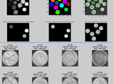
Perfect for the beginner, this demo illustrates simple object detection (segmentation, feature extraction), measurement, and filtering. Requires the Image Processing Toolbox (IPT) because it demonstrates some functions supplied by that toolbox, plus it uses the "coins" demo image supplied with that toolbox. If you have the IPT (you can check by typing ver on the command line), you should be able to run this demo code simply by copying and pasting this code into a new editor window, and then clicking the green "run" triangle on the toolbar.
First finds all the objects, then filters results to pick out objects of certain sizes. The basic concepts of thresholding, labeling, and regionprops are demonstrated with a simple example.
It's a good tutorial for those users new to MATLAB's image processing capabilities to learn on, before they go on to more sophisticated algorithms.
Tested with R2008b through R2011b.
Image Analyst (2021). Image Segmentation Tutorial (https://www.mathworks.com/matlabcentral/fileexchange/25157-image-segmentation-tutorial), MATLAB Central File Exchange. Retrieved .
Inspired: Cell_Analyzer, SimpleColorDetectionByHue(), Image segmentation using fast fuzzy c-means clusering, M-code for leaf identification
Find the treasures in MATLAB Central and discover how the community can help you!
Start Hunting!Create scripts with code, output, and formatted text in a single executable document.
If I have my own images, how to put them in the code? Does the code find the center mass of each image?
Contact for code and support mycodeworklab@gmail.com
Whatsapp +91 9464894829
1. RGB-Image-encryption-based-on-chaotic-system-and-DNA-algorithm
2. Image-encryption-based-on-chaotic-system
3. Image encryption by generating Halton sequences
4. Chaos-based-Image-Encryption
5. Text-Encryption Matlab code for AES,DES,Hybrid AES-DES and AES w/ chaos
6. Image encryption and decryption using chaotic key sequence generated by sequence of logistic map and sequence of states of Linear Feedback Shift Register
7. A New Approach of Image Encryption Using 3D Chaotic Map to Enhance Security of Multimedia Component
8. MATLAB was used for the implementation of Chaotic Digital Image Encryption.
9. Multiple-Color-Image-Encryption
10. Cryptanalyzing an Image Scrambling Encryption Algorithm of Pixel Bits.
11. Novel Image Compression encryption hybrid algorithm based on a key-controlled measurement matrix in compressive sensing.
12. An Image Encryption Scheme Based on a Hybrid Model of DNA Computing, Chaotic Systems and Hash Functions.
13. Colour image encryption algorithm combining Arnold map, DNA sequence operation, and a Mandelbrot set.
14. Advanced Encryption Standard
15. Reversible-Data-Hiding-by-Reserving-Room-Before-Encryption-MATLAB.
16. Image Encryption and Decryption Using Logistic Map Equation and Linear Feedback Shift.
17. A simple Matlab implementation of the algorithm presented in the paper: "Reversible-data-hiding-in-Encrypted-image"
17. Image encryption and encoding methods
19. DES 64bit Encryption and Decryption
20. Matlab project on blind digital watermarking and encryption.
21. Encrypting an image using Salient Object Detection and K-Means Clustering.
22. Recurrent Scale Approximation for Object Detection in CNN.
23. Object detection via a multi-region & semantic segmentation-aware CNN model.
24. R-FCN: Object Detection via Region-based Fully Convolutional Networks
25. Adversarial Examples for Semantic Segmentation and Object Detection.
26. Object Detection in Videos toolkit for VisDrone2019
27. Computational biology and medical image processing scripts and programs.
28. A MATLAB library/toolbox providing access to image registration suitable for use with medical images.
29. Lung medical image analysis and visualisation software for Matlab.
30. Medical ultrasound image processing.Carotid ultrasoung segmentation using RF data.
31. A Phase Congruency and Local Laplacian Energy Based Multi-Modality Medical Image Fusion Method in NSCT Domain.
32. Automatic tool for landmark localisation in 3D medical images.
33. Advanced-Medical-Image-Processing.
34. Recognizing and Refining the location of Individual Vessels in Segmented Retinal Images.
35. Image segmentation method on medical image is provided and tested.
36. 3D non-rigid image registration for medical and synthetic images using truncated hierarchical B-splines (THB-Splines).
37. Laplacian Re-Decomposition for Multimodal Medical Image Fusion[J]. IEEE Transactions on Instrumentation and Measurement, 2020.
38. Medical software for Processing multi-Parametric images Pipelines.
39. Prostate cancer segmentation based on MRI and PET images.
40. Image segmentation methods for biomedical purposes such as cell segmentation, blood vessel segmentation (eye blood vessels), and segmentation of brain tumors.
41. Medical Image Analysis Breast Cancer Lesion Detection.
42. Medical image enhancement based on nonlinear technique and logarithmic transform coefficient histogram matching.
43. Machine Learning: A Bayesian and Optimization Perspective.
Your tutorials and answers are very helpful. I like it
"Attempt to execute SCRIPT regionprops as a function:
E:\Programy\MatLab\toolbox\images\images\regionprops.m
Error in BlobsDemo (line 140)
blobMeasurements = regionprops(labeledImage, originalImage, 'all'); "
Incredibly helpful
Respected Image Analyst, Your tutorials and answers are very helpful. Every time it saves me. Sir I am looking for code for below algorithms (any one/two will be fine)
1. Improved Local Ternary Pattern.
2. Elongated Local Ternary Pattern.
3. Elongated Local Quinary Pattern.
4. Ensembled of Local Quinary Pattern.
can you please help me aboutthis.
Thanks.
can anyone help me out in list of statistical parameters obtained from a segmented image?
Dear Image Analyst, Your tutorial is very useful. As a beginner to image analysis, it helped me understand how to segregate relevant sections of my images and analyze my data. I used your script as part of my data analysis. Please let me know how I can reference your code in my publication.Thanks for your help!
safina, image processing algorithms need to be adapted to the images. Whether it will work on your specific coins is just something you have to test.
vv_art, if you have questions about specific image processing algorithms, ask them in the Answers forum, not here. There we can give you a proper answer, something I can't do in the brief comment section here. Thanks.
dear sir.
Currently i am working on detection of different colors in a resister using image processing.But, I am unable to do this for all images and all colour detection.Please help how to approch.if possible please send me the code sir.
Thank you
Could we run this code for different images of coins? Surely it will run on them?? Does Quality of picture matter?
Thanks alot! segmenting by thresholding dark regions was surprisingly hard to track down online but yours works like a charm!
Thank you so much this is really great, i want to know how to apply this to an image ?
Thank you so much! How would i apply this to a video?
Dear Image Analyst,
Your tutorial on image segmentation was a great help. I have ran into a following problem and wonder whether you can guide me. I have a segmented image which contains a part of the rock which consisted the fractured area and also the white corner regions. I want to calculate the total no of white pixels within the dark region (i.e no of fractured pixels). How can I do that avoiding the corner white areas.Much appreciated.
Svetlana, look in the Answers forum for "Spatial calibration" because I've posted code for converting pixels into real world units such as mm.
Hello,
The area you get are in units pixels ... I'm using a microCt scout image and need to find areas of similar shapes but in mm units. Do you have an idea of how I can achieve this?
Hi image Analyst-this code was really helpful. Anyway, i am currently working on my final year project, and i am using your coding to detect normal cell and sickle cell. The coding work perfectly, however, it didn't detect the sickle cell. May i know what is your parameter value? and which part should i adjust the value of the parameter
Hi Image Analyst - this code is really very helpful. thanx
plz clarify me one thing that for the purpose of image segmentation, if we use matlab function ( regionprops) then is this any algorithm? or just a function
Hi Image Analyst - I thought this might be the case. How would one change the pixel values to black or white?
Many thanks for your help, Joe
hello sir,
can we save the different coins separately into a folder??if yes,Can you explain how with a code
Joe Perkins, images must remain rectangular so the coins MUST have some background outside the round regions of the coin itself. All you can do is to change the background, not remove it. You can have this either the original background, or some uniform intensity like black, white, or some gray level.
Can this script be used to count black or white pixels of an image? I was trying to identify if I could do that by looking at the code but I'm not sure due to the complexity of this example.
brilliant
Hi Image Analyst,
Brilliant demo thank-you!
How would you go about separating the coins in the final images from the background around them so that the output images contain the coins only?
Many thanks,
Joe
Dear Image analyst,
Thank you very much for the codes again. I am a neuroscientist from MIT and I'm really impressed by your recent code. I have an example neural calcium imaging tiff file (stack a tiff files from one video). I have to extract calcium signals (peaks, ROIs) from individual synaptic site. The goal is to map the synapses when they are active. I really believe that your code can solve of my problem. Could you please help more in this regard?
Thank you so much,
Kind regards
Suresh
I really enjoyed this code. I have some back ground in image analysis yet these codes are very informative.
Please post more advance example as well. Thank you
Good demon to novice like me. Thanks alot
Hello Image Analyst, I am going to do some analysis on an image and i need to write a code to recognize the coordinates of different points of red color automatically without human interfering. I will be grateful if you propose some methods. Thanks in advance.
Nice job.Thanks for your sharing.
Nice tutorial. Thank you
sairam to all members ,
can anyone give me the code of otsu thresolding for applying on to thyroid dicom images
Excellent!
I did it the hard way until I saw this and then it was so-easy.
Thanks
GUD
Awesome work. Inspired by you I have also uploaded 13 of my projects for sharing knowledge. Plz checkout https://in.mathworks.com/matlabcentral/fileexchange/?term=authorid%3A620308&sort=ratings_desc
anyone help me? In my thesis work on content image retrieval using HSV CCV and LBP ..please provide me email id if possible mine is himanshu.atra@gmail.com
Quite helpful. And so nicely documented.
Hello, I currently have an image of the veins binarized. I've tried using parts of your code so that i could then find the coordinates of the the edges/nodes of the image but have had no luck. Any tips would be appreciated.
If anyone has specific questions about how to adapt this demo to their specific image, post the image and problem description to the Answers forum here on the Mathworks website.
hello dear i need your kind support to find the center of an object in a binary image in order to i can crop that object from the binary image, note that the binary image has only one object no more so that ow can i determine the center of that object
Thanks, the file helped me to understand segmentation.
How can same be used with the DICOM images? Can you help me with the feature extraction of the images?
Wow; I agree you are the best teach at Image analysis and processing; This tutorial helped me a lot indeed THANK YOU VERY MUCH;
I would like to learn from you more as I also approach my project; Please write to me at: powellorenz@gmail.com
So I can ask you some questions.
THANKS
thanks for your sharing, this is helping me
hye. can i ask u some private question on how i can detect the circle like u do but my circle not really circle because the picture i snap was a tennis ball and pingpong ball.can u give me a solution please.i try your coding but it didn't seem work.really need your help.and please email me at fatinsarah31@gmail.com if u feel free.tyvm
Excellent, thank you
Ramses, you just flip the sign of the > sign to < so that it finds dark objects instead of bright objects.
How would one handle images where the object intensities are much lower, say segmentation of organs in a CT?
Cheers,
Ramses H.
Thank you, this is excellent.
its very useful for me.. thank you..
Thank you for the amazing tutorial. what do i do if i have to extract a certain section from image who's orientation can be anyway.
I have a question,
is there an automatic way to filter the unwanted regions from a binary image , where the wanted regions are character candidates in different scales (sometimes big letters and sometimes small)?
please help me out
important
Perfect!!! thank you for sharing.
great work !!
thank you a lot.
please send us the code
thank for sharing
Thanks a lot for sharing ;)
Nice work!
Dear Image Analyst,
Thank you for this beautiful code. I'm trying to use it for oil palm ripeness detection.
Looks simple and sophisticated at the same time! Full of comments and as such easy to follow the chain of thoughts of the author. One can simply make adjustments based on his/her needs. Great job Image Analyst!
hi
i get an error...please help me.
??? Error using ==> cd
Cannot CD to (Name is nonexistent or not a directory).
Dear Image Analyst,
Thanks for this beautiful code. I have a minor question concerning about the 'keeper' blob image. Is that possible that we segment the keeper blobs while still keep their numbers on it? Like tracing which number of blobs are the brightest and which number are the darkest? How should I write the code?
Thanks for your help!
Hello,
I used this code to find the Area of the each object this file does,But I am in need of number occurrence of same object should be known and that should be plotted..
Works perfectly - very helpful!!
I also got the same errors as xsfeng when I run it.
Error using fileparts
Too many output arguments.
Error in BlobsDemo (line 191)
[thisFolder, baseFileName, ext, version] = fileparts(thisFile);
dghgfh
Dear Image Analyst,
I got the error information when I run this scripts:
Error using fileparts
Too many output arguments.
Error in BlobsDemo (line 191)
[thisFolder, baseFileName, ext, version] = fileparts(thisFile);
Could you please help me with this? Many thx!
ok good programing
mustafs, the more sophisticated algorithms are more dependent on image content. More complicated content requires more complicated algorithms. I did add some demos on color segmentation, but I want to stick to fairly standard kinds of segmentation since there are an infinite number of unique, peculiar ones.
dear image analyst,
can you please make to us more advance segmentation algorithms for different industrial application
the segmentation process is the most hard operation for all
Hello image analyst,
How did you obtain the values to divide between foreground and background, at your thresholding code.
for example, why 70 and 0.94
thresholdValue - 70), double(0.94 * maxYValue(2))
Looking at the code taught me a few new tricks ...very nice.
Awesome, a nice presentation on feature extraction.
This is so helpful. Thank you so much!
Excellent!!!
Nina, to do that exactly, you'd need to use John D'Errico's interparc program. Look for it in the File Exchange. You can also subsample, like thisBoundary(1:10:end,2) though these won't necessarily be evenly spaced in distance.
Very useful demo. I do have a question Image Analyst - I hope you see this! For the part of the code:
boundaries = bwboundaries(binaryImage);
numberOfBoundaries = size(boundaries);
for k = 1 : numberOfBoundaries
thisBoundary = boundaries{k};
plot(thisBoundary(:,2), thisBoundary(:,1), 'g', 'LineWidth', 2);
end
hold off;
Is it possible to plot say only 10 points equally spaced around the boundary? Are they in order say clockwise or anticlockwise?
Very nice, saved me at least one day!
is the actual tutorial available to watch online? cannot seem to locate it.
If anyone has any questions about their own specific images, they should ask in the Answers forum (look at the menu above). Do not put your specific question here as a comment on this program. I can help there in Answers. This page is not meant to be a discussion forum.
Hello Image Analyst ,
your demo is excatly what I am looking for to analyse my fluorescence images. However my images are taken with a 12 bit peltier cooled fluorescence camera and saved as .tif. I can hence not display their histograms without getting an error:
" Warning: Image is too big to fit on screen; displaying at 67% "
I tried to plot the histogram using
plot_size = get(0,'ScreenSize');
fg1 = figure(1);
set(fg8, 'Color', [1 1 1], 'Position', plot_size, 'Visible', 'on');
[pixelCount grayLevels] = imhist(orginalimage);
bar(pixelCount); title('Histogram of orginalimage');
xlim([0 grayLevels(end)]);
but it didnt help. I would really appreciate any help and comments.
Cheers
microF
Hey this demo has been very useful.....my project is wavelength calculation using diffraction grating, so we get an image which will be a series of round dots and we should find the distance between two successive points.......so with this blob i can find the dots but how to find the centres of those dots?????? plz help me anyone... i would be greatful......ty in advance
Thanks for showing us a basic demo. It helped a lot!
thanks for sharing this demo. This provides a very good example for beginners.
Christie, when you adapted it, you had it read in a color image, even if it doesn't look like it. Do this to prove: [rows columns NumberOfColorChannels] = size(yourImage). Use rgb2gray() right after you call imread() to convert your color image into a gray scale image.
Hey, Image Analyst,
Sorry, I tried to send this as a message first but kept getting an error from the site. Anyway, I am using your BlobsDemo to find the area of leaf surfaces traced onto a piece of graph paper. The code has no problem translating it into a binary image with clearly defined leaves and no graph paper grid, but when it tries to label each blob I get the following error:
??? Error using ==> iptcheckinput
Function BWLABEL expected its first input, BW, to
be two-dimensional.
Error in ==> bwlabel at 67
iptcheckinput(BW, {'logical' 'numeric'}, {'real',
'2d', 'nonsparse'}, ...
Error in ==> LeafArea at 48
labeledImage = bwlabel(binaryImage, 8); % Label
each blob so we can make measurements of it
What does it mean when it says the image isn't 2-dimensional?
Thanks for your help,
Christie
I'm finding your Demo quite very useful in my work. Thank you.
it's nice info for beginner thank's
Syahrul, it does do segmentation into several areas - each coin is a different area. How can you label something without having a binary image? You can't, but why would you want to avoid labeling even if you could?
sivan, you must have a color image for some reason. You probably changed the filename to one of your images. Coins.png that my demo uses is a monochrome (gray scale) image.
is it possible to do segmentation into several areas, not binarization before labeling?
If anyone wants to ask me a question, click my name above, then click the "Contact Author" link.
can any wan help me...why i got error in the code when i try to run it..in line 41-[pixelCount grayLevels] = imhist(originalImage);...this code..the error said
("??? Error using ==> iptcheckinput
Function IMHIST expected its first input, I or X, to be two-dimensional.")
why it said so...i just keep on follow the code given but suddenly it give error
Hi I appreciate this demo very much. I modify your code to work along with watershed segmentation for my image (blood cell), and it turns out good. However, how do I filter the background after I plot the boundaries of the selected cell? (so that the resulting image only shows the blobs without the background) The cropping works very well too but I don't want to involve the background. Thank you.
German:
You ask this kind of question on the newsgroup or on Answers, not as a comment on people's File Exchange submissions. I'd be happy to answer it there.
Please I need some help !!! I need to capture a basic color card with my webcam, then I need to determine what color is it. Matlab code analize and gives the answer. Example: The card is yellow. Can someone of you Help me please with the MatLab code ???? TKS A lot !!!
Hi guys !!! I need to capture a primary color card with my webcam, then I need to determine what color is it ??? Can someone of you Help me please with the MatLab code ???? TKS A lot !!!
Thanks!
I should read code comments more thoroughly. No need to answer. Thanks!
thank you
Please can somebody send me the matlab code for gray scale image segmentation plzzzz...?I will be very greatful
Hello!
I'm pretty new to matlab.Can you offer some help regarding my issue?
I need a code to extract objects from a single image and write the extracted objects to separate images.As an example if an image consists of say an elephant and tree , I want to extract these two objects and write as separate images..
Very cool code.
Manhal, I used Photoshop to create an image with 12 coins and it worked just fine. You must have done something wrong.
Vivek, what is it about this that made you think it was a 1 star? Is there anything I could improve with it to help beginners understand it better?
and one more thing, I tried to change the image that is used in the BlobsDemo, by adding 2 more coins, but still show that we already have 10 objects...
is there something should I change to make it works??
Useful and well-commented code that demonstrates image thresholding. Connected components, including real world objects, in an image is of interest to many and this code provides the starting point for such projects. Thanks IA.
An excellent demonstration of simple image segmentation. Also a nice introduction to the image toolbox in Matlab. The functions for dealing with connected components (centroid, mean, etc) should be extremely useful.
J.D, you need to get some model of skin color. I know that it's a boomerang- or banana-shaped cluster in HSV color space. You need to define parameters to model that, and then segment it, possibly with a 3D lookup table instead of a mathematical model, or with the mathematical model if you can come up with the formulas. But I know you can't simply do high and low thresholding in any color space to carve out a box that selects the skin color pixels.
hello ~ i juz want to ask you what should i do if i want to recognize color of human skin~ i want to get its hsv value for every pixel of the human face ~ and then exlude the eye and mouth part~ i want to get the value as my input to train in neural network~ can u help me~ urgent.. tq~
Christopher. Yes there are lots of more complicated images that could be used that would be more challenging. I work with many of those kinds of images. However I know from my newsgroup postings that even code as simple as this blows a lot of people away. So thanks for the suggestion but I think I'll keep it simple. I do have some color demos posted that are slightly more challenging.
Hi I am working in my master about breast cancer mass detection the first step is to determine region of interest which is the breast contour and i need help in this step as i don't know how to do this so please if there is anyone who can help me
thanks
i am just trying it
Interesting demonstration ImageAnalyst. Fortunately the 'coins' sample image has high contrasting content which allows for easy selection of the threshold. Plus the coins allow for the 'imfill' operation to perform the majority of the work.
It would be interesting to see results on an image with a much smoother histogram but this is where image segmentation gets complicated I guess...
All in all, a nice demonstration of the capabilities that both the IPT and Mathematical Morphology can provide in image processing applications.
hello every body, can you give me a code for building extraction using mathematical morphologie ?? and thanks a lot
Hi, I've tried your demo but it's not working for 3D images. Is there any way to solve this problem? Thanks
Like other submissions that are meant to demonstrate certain toolboxes, you need to have the toolbox. If you don't have it, you can buy it, or write the necessary toolbox functions yourself. Generally they are the functions starting with the letters "im" or "bw".
What if I don't have the IPT??
I just realized I use Matalb R2007b, not R2008. I guess that is the problem. Sorry about that.
Very useful...great work...
great work..I have one question for U. using my web cam i will capture a picture which will contain a ball in a room. now how can i separate the ball (circular shaped) from the background and i want to find the area of that ball(circle). how can i edit your code for this particular task.
Jimmy - ask in the newsgroup. Let's take this conversation there.
Ok let's forget about GUI, I am not even sure if I need to use it. The reason I asked if I can get more than one image read is than after I modified the code I got my the centroids of all objects in the images. The results are in vector form [x y]. For example
[29 134]. I want to make a loop going through all images and objects in them, reading those measurements. Can you give me an advice? Thank you
Excellent!!!
Jimmy, just paste whatever code you want into line 1076 of that app after you've deleted lines 1076-1164. Then just run it with the green triangle. You need all the rest of the code because that is the GUI. To select images, you simply click on their names in the listbox like you'd do with any app. No code is needed from you to handle clicking on a listbox - that's already done for you. Just click on the images you want to process and click the analyze button and it will analyze only those that you've clicked on.
Ok I did as you said Image Analyst.I got some errors. Now do I only use 1076 - 1164 code or the whole Magic code which is huge? And another thing how do I select images from a folder? Do I need to put that into the code? Thank you
Jimmy, just follow these easy step by step instructions.
1. Open both his MAGIC code and my BlobsDemo code in MATLAB.
2. Copy all of my BlobsDemo code into the clipboard.
3. Go to AnalyzeSingleImage in his code, currently line 1075, and simply delete all of that function, which means delete lines 1076 - 1164. This is just like it says to do in the comments on line 1070 and 1071 of his code.
4. Now simply paste in my BlobsDemo code.
Now it should work. Adapt my/your code in that function as you see fit.
Thank you for your reply Image Analyst. Yes, I know that I need to make changes according to the pictures regarding threshold etc.. What I don't understand is how to plug that code into AnalyzeSingleImage(). Is it possible for you to send me the example on testbest1@yahoo.com. Thank you
Jimmy, sure - just copy the code into your own program. One way to do it might be to use this in conjunction with MAGIC (http://www.mathworks.com/matlabcentral/fileexchange/24224) which does batch processing. Just take this code and plug it into his AnalyzeSingleImage() function. Then you'll be able to select several images from your folder, and process them. Of course, my code has several things that are special for the coins image and you'd have to modify several things for your images, such as the thresholds, etc.
Great Work Image Analyst, very useful indeed. I want to ask one question: is there any chance that it can be applied to more than one image simultaniously? And if there is how can it be done. Thank you
Thomas, this is not an error in my code. sprint() is not a function - ParseInputs() should have called sprintf() instead. regionprops() and ParseInputs() are Mathworks code, not mine. I searched the R2008b version of ParseInputs and it no longer has sprint() in it, so they've fixed that bug.
Sorry that I can't test this with every prior version of MATLAB. I know it definitely works with R2008a and later. Since you are several years behind I suggest you get the most modern version of MATLAB.
Not working for me:
Running BlobsDemo.m...
??? Undefined command/function 'sprint'.
Error in ==> regionprops>ParseInputs at 878
msg = sprint('This measurement is not a string: "%d".', list{k});
Error in ==> regionprops at 114
[L, requestedStats] = ParseInputs(officialStats, varargin{:});
I got :
MATLAB Version 7.1.0.246 (R14) Service Pack 3
and the IPT
an excellent and exceptionally well commented image processing demo by a seasoned ML user and senior CSSM citizen...
a few changes i'd make:
- put it into a function
-> no need for the bad CLEAR ALL...
- make the figure size smaller -and-
- put an ECHO ON/OFF at the beginning/end of the demo
-> for users to be able to enjoy the comments during run-time...
just a few thoughts
us