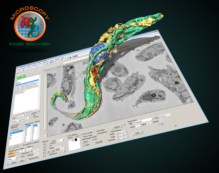
Microscopy Image Browser (MIB)
MIB is a package for segmentation of multi-dimensional (2D-4D) microscopy datasets

You are now following this Submission
- You will see updates in your followed content feed
- You may receive emails, depending on your communication preferences
Cite As
Ilya Belevich (2026). Microscopy Image Browser (MIB) (https://github.com/Ajaxels/MIB), GitHub. Retrieved .
Acknowledgements
Inspired by: view3d.m, Local Normalization, extrema.m, extrema2.m, IMAGEVIEWER, findjobj - find java handles of Matlab graphic objects, 3D Euclidean Distance Transform for Variable Data Aspect Ratio, Region Adjacency Graph (RAG), stlwrite - write ASCII or Binary STL files, maxflow, Viewer3D, export_fig, Hessian based Frangi Vesselness filter, Accurate Fast Marching, Fast/Robust Template Matching, imgaussian, Image Edge Enhancing Coherence Filter Toolbox, Highly portable JSON-input parser, Jann5s/measuretool, Render RGB text over RGB or Grayscale Image, improved xlswrite.m, xml2struct, struct2xml, IMCLIPBOARD, Hardware accelerated 3D viewer for MATLAB, mdrohmann/mtocpp, drifty_shifty_deluxe.m, regionprops3
General Information
- Version 1.302.0 (42.7 MB)
-
 View License on GitHub
MATLAB Release Compatibility
- Compatible with any release
Platform Compatibility
- Windows
- macOS
- Linux
| Version | Published | Release Notes | Action |
|---|---|---|---|
| 1.302.0 | Added reference to MIB2, which is recommended to use instead of MIB for Matlab R2014b and later |

